
Study of Representative Plant (Epidermal cells of Rhoeo sp ) by Microscopy
Aim –
To prepare stained temporary mount of leaf peel (Rhoeo plant leaf)
Materials Required –
To watch a Rhoeo leaf under microscope we use those below mentioned materials.
- A potted plant (Rhoeo species)
- Watch Glass
- Forceps
- Dropper
- A compound Microscope
Procedure of Experiment –
- A healthy Rhoeo leaf from a potted plant is taken.
- A part of the peel was removed with the help of forceps from the lower surface of the leaf.
- The peel was then placed in a watch glass, which contain water in it.
- With the help of forceps the peel was placed on a clean glass slide.
- Lastly the slide was observed under the microscope.
Observation –
Following are the observation, after visualization of this prepared slide under the microscope.
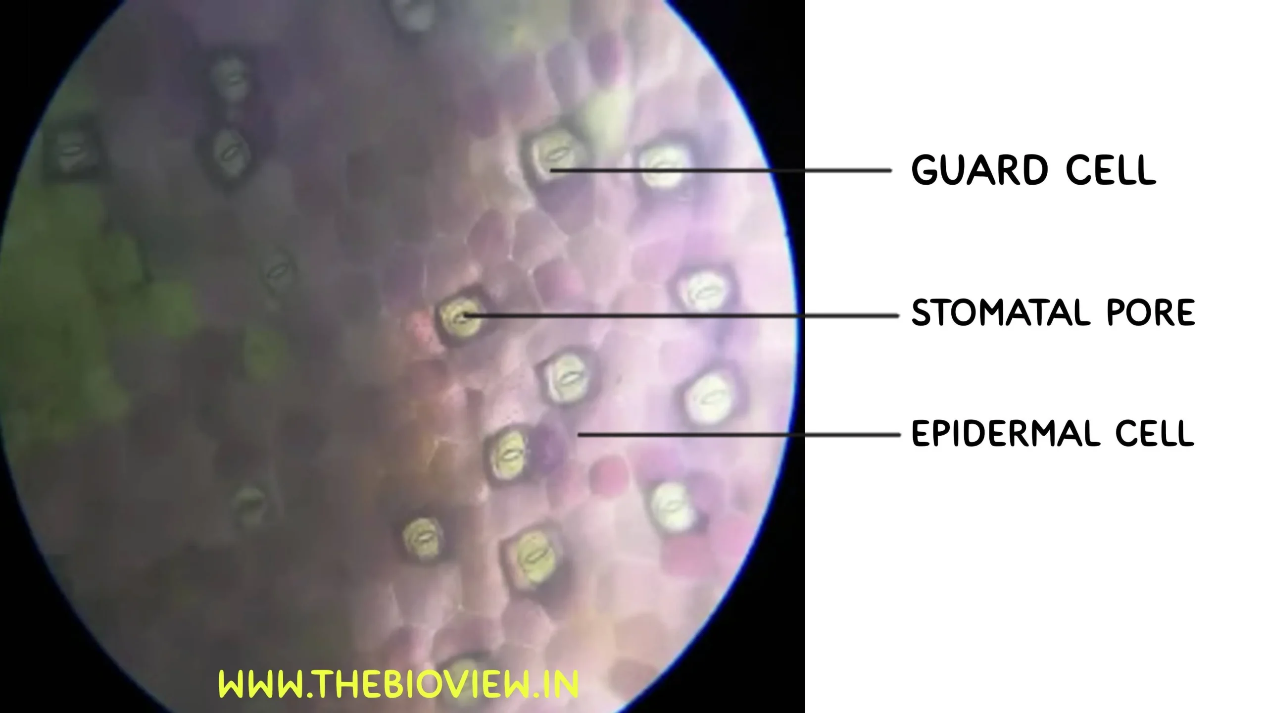
- The epidermal cells are visible. They are irregular in outline (more or less cuboidal with no intercellular space).
- Many small pores (stomata) are seen scattered among the Epidermal cells (Lower Epidermis).
- Each pore is guarded by two bean shaped Guard cells.
- Subsidiary cells surrounding the guard cells were also visible.
- Stomata are present in the Epidermal Cells of the lower surface of the leaf.

Precautions –
We always need to folow some basic precautions in the laboratory while we are working in any experiment. For this Rhoeo leaf experiment we need to care about the precaution that
- The leaf peel should be cut to a proper size and folding or curling should be avoided.
- Proper microscopic observation should be done carefully.
Theory –
The cells on the surface of a leaf, the Epidermal cells form the barrier between air or water and the inner cells of the leaf. The surface may have a wax coating or hairs as protective structures. Stomata are specialized structures found on the Epidermal surface of the leaves of plants that are used to control exchange of gases during Photosynthesis and Respiration. Each stomata has a slit like opening called Stomatal pore, which is surrounded by a pair of specialized cells called Guard cells that are responsible for regulating the size of the opening. Guard cells contain chloroplasts that swell or shrink in association with osmotic changes. When guard cells are turgid , a space between the cells opens – the Stoma or Stomatal pore through which CO2 (carbon dioxide) enters the plant and Oxygen (O2) and water (H20) is released through the Stomatal pore into the atmosphere in the form of water vapour through the process called Transpiration . Plants need to regulate the amount of time the pore is open to maximize carbon dioxide (CO2) entry for photosynthesis and limit water (H20) loss (transportation) keeping between the epidermal layers are Mesophyll cells , which carry most of the chloroplast and where photosynthesis occur.
This method describes how to prepare a peel of a leaf epidermis for microscopic observation of the Epidermal cells, guard cells and leaf hairs if they are present. Plants vary in the shape of Epidermal cells and size and number of stomata on each surface. Usually the lower surface of a dicot leaf has a greater number of stomata which in monocot leaf is more or less equal in both surfaces.
Conclusion –
Hope you are now well aware about the rhoeo leaf experiment and How to see epidermal cells of Rhoeo plant. This was the practical named Study of a representative plant cell by Microscopy.