Aim :
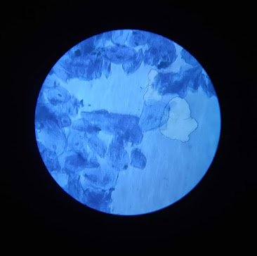
To Prepare Stained temporary mount of human cheek cells.
Theory –
The body of all animals including humans is composed of cells. Unlike Plant cells, animal cells don’t have cell wall. The outermost covering of an animal cell is a Plasma membrane. A cell membrane is a semipermeable membrane that surrounds the cytoplasm. The cytoplasm, nucleus and other cell organelles are enclosed within the cell membrane. The cytoplasm in animal cell is denser, granular, and occupies a large Space. Epithelial tissue is the outermost covering of most organs and cavities of animal body. The vacuoles in animal cell is smaller in size or absent. The nucleus is Present at the centre of the cytoplasm. The absense of a cell wall and a prominent vacuole are indicators that help in identifying animal cells, such as cells seen in human squamous epithelial cells. This cells are polygonal in shape and arranged compactly in a single layered without any intercellular matrix.
Instruments Required –
1. Glass slide and cover slip.
2. Toothpick
3. Methylene Blue
4. Watch Glass
5. Distilled water
6. Dropper
7. Blotting Paper
8. Compound Microscope
Procedure –
1. The inner side of the cheek was gently scraped using a toothpick, which will collect some cheek cells.
2. The cells were placed on a glass slide that has a drop of distilled water in it.
3. The Water and the cheek cells were mixed using a needle.
4. A few drops of methylene blue solution was taken using a dropper and added to the above mixture on the slide .
5. After 2-3 minutes any excess water and stain was removed from the slide using a blotting Paper.
6. A clean cover slip was carefully placed on the mixture with the aid of needle.
7. Using a needle the coverslip was gently pressed to spread the epithelial cells.
8. Any extra liquid around the cover slip was removed using a blotting paper.
9. Lastly, the glass slide was placed on the stage of the Compound microscope and viewed.
Observation –
1) A large number of flat and irregular shaped cells were visualised. These cells are small and compactly arranged to form a continuous layer.
2) The cells don’t have cell wall. However, each cell has a thin cell membrane.
3) A deeply stained nucleus is observed at the centre of each cell.
4) No Prominent vacuoles were observed in the cells.
Read more –
Conclusion –
As the cells observed don’t have a cell wall, not a Prominent Vacuole, the cells of the specimen on the slide were of animal cells.
Precaution –
Ensure that the toothpick used to scrap the squamous is clean so that it does not cause any infection to the cheeks.

